")
Unlocking the Invisible: How X-ray Microtomography is Transforming Material and Biological Analysis. Discover the Science, Technology, and Future Impact of High-Resolution 3D Imaging. (2025)
- Introduction to X-ray Microtomography: Principles and Evolution
- Core Technologies: Hardware, Software, and Imaging Techniques
- Key Applications in Materials Science and Engineering
- Breakthroughs in Biological and Medical Research
- Comparative Analysis: Micro-CT vs. Other Imaging Modalities
- Industry Leaders and Notable Systems (e.g., bruker.com, zeiss.com)
- Data Processing, Visualization, and Interpretation Challenges
- Market Growth and Adoption Trends: 2024–2030 Forecasts
- Emerging Innovations and Future Directions
- Ethical, Regulatory, and Accessibility Considerations
- Sources & References
Introduction to X-ray Microtomography: Principles and Evolution
X-ray microtomography, often abbreviated as micro-CT, is a non-destructive imaging technique that enables the visualization and analysis of the internal structure of objects at micrometer-scale resolution. The fundamental principle of X-ray microtomography is based on the differential absorption of X-rays as they pass through a specimen. By acquiring a series of two-dimensional radiographic projections at different angles, computational algorithms reconstruct a three-dimensional representation of the object’s internal features. This approach is analogous to the medical computed tomography (CT) used in clinical diagnostics, but micro-CT achieves much higher spatial resolution, making it invaluable for research in materials science, biology, geology, and industrial quality control.
The origins of X-ray microtomography can be traced back to the broader development of X-ray imaging following Wilhelm Röntgen’s discovery of X-rays in 1895. The first clinical CT scanner was introduced in the early 1970s, revolutionizing medical diagnostics. However, the need for higher resolution imaging in non-medical fields led to the adaptation and miniaturization of CT technology. By the late 1980s and early 1990s, advances in X-ray sources, detector technology, and computational power enabled the emergence of micro-CT systems capable of resolving features down to a few micrometers. These systems were initially developed in academic and government research laboratories, such as those affiliated with the National Institute of Standards and Technology and the European Synchrotron Radiation Facility, which played pivotal roles in advancing X-ray imaging techniques.
Modern X-ray microtomography systems utilize either laboratory-based microfocus X-ray tubes or high-brilliance synchrotron radiation sources. Synchrotron-based micro-CT, available at large-scale facilities like the European Synchrotron Radiation Facility, offers exceptional spatial and temporal resolution, enabling dynamic studies and phase-contrast imaging. Laboratory micro-CT instruments, meanwhile, have become increasingly accessible and are widely used in academic, industrial, and clinical research settings. The National Institute of Standards and Technology and similar organizations have contributed to the development of calibration standards and best practices, ensuring the reliability and reproducibility of micro-CT measurements.
Over the past three decades, X-ray microtomography has evolved from a specialized research tool to a mainstream technique with broad applications. Its ability to provide detailed, three-dimensional, and non-destructive insights into the internal architecture of materials and biological specimens continues to drive innovation in fields ranging from biomaterials and paleontology to electronics and additive manufacturing.
Core Technologies: Hardware, Software, and Imaging Techniques
X-ray microtomography (micro-CT) is a non-destructive imaging technique that enables the visualization and quantitative analysis of internal structures in objects at micrometer-scale resolution. The core technologies underpinning micro-CT systems encompass advanced hardware, sophisticated software, and specialized imaging techniques, each contributing to the method’s precision and versatility.
Hardware forms the backbone of micro-CT systems. The primary components include a high-brightness X-ray source, a precision sample stage, and a high-resolution detector. Modern micro-CT scanners utilize microfocus X-ray tubes capable of producing spot sizes down to a few micrometers, which is essential for achieving high spatial resolution. The sample stage is engineered for sub-micron positioning accuracy, allowing for precise rotation and alignment during scanning. Detectors, often based on scintillator-coupled charge-coupled devices (CCDs) or complementary metal-oxide-semiconductor (CMOS) technology, convert X-rays into visible light and then into digital signals, enabling the capture of detailed projection images. Leading manufacturers such as Carl Zeiss AG and Bruker Corporation have developed micro-CT systems that integrate these hardware elements to deliver robust and reproducible imaging performance.
Software is critical for both the control of hardware and the reconstruction of three-dimensional datasets. Acquisition software manages the synchronization of X-ray emission, sample rotation, and image capture, ensuring optimal data quality. Reconstruction algorithms, such as filtered back projection and iterative reconstruction, transform the two-dimensional radiographic projections into volumetric datasets. Advanced software platforms also provide tools for image segmentation, quantitative analysis (e.g., porosity, density, and morphometry), and visualization. Companies like Thermo Fisher Scientific and Carl Zeiss AG offer proprietary software suites that support a wide range of analytical workflows, from materials science to life sciences.
- Imaging techniques in micro-CT have evolved to address diverse application needs. Standard absorption-contrast imaging remains the most common, relying on differences in X-ray attenuation to reveal internal features. Phase-contrast imaging, which exploits variations in the refractive index, enhances the visibility of low-density or weakly absorbing materials, making it valuable in biological and polymer research. Dual-energy and multi-energy micro-CT techniques enable material discrimination by acquiring images at different X-ray energies, facilitating compositional analysis. Innovations in in situ and time-resolved micro-CT allow researchers to observe dynamic processes, such as deformation or fluid flow, within samples under controlled environmental conditions.
Together, these core technologies—hardware, software, and imaging techniques—define the capabilities and expanding applications of X-ray microtomography in 2025, supporting research and quality control across materials science, biomedical engineering, geology, and beyond.
Key Applications in Materials Science and Engineering
X-ray microtomography (micro-CT) has become an indispensable tool in materials science and engineering, offering non-destructive, three-dimensional imaging at micrometer-scale resolution. This technique enables researchers to visualize and quantify internal structures, defects, and compositional variations within a wide range of materials, from metals and ceramics to polymers and composites.
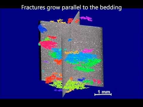
One of the primary applications of X-ray microtomography in materials science is the characterization of microstructure. By providing detailed 3D images, micro-CT allows for the analysis of grain boundaries, phase distributions, and porosity in metals and alloys. This is crucial for understanding mechanical properties and failure mechanisms, as well as for optimizing processing techniques. For example, in additive manufacturing, micro-CT is used to detect internal voids, cracks, and inclusions that may compromise the integrity of printed components. Such insights are vital for quality assurance and for advancing the development of new materials with tailored properties.
In the field of composites, micro-CT enables the visualization of fiber orientation, distribution, and the presence of voids or delaminations. This information is essential for predicting the mechanical performance of composite structures and for improving manufacturing processes. Similarly, in ceramics and porous materials, micro-CT is used to assess pore size distribution, connectivity, and tortuosity, which are key parameters influencing properties such as permeability and strength.
Another significant application is in the study of degradation and failure processes. Micro-CT can monitor the evolution of cracks, corrosion, and other forms of damage in situ, under mechanical or environmental loading. This capability supports the development of more durable materials and the design of components with enhanced lifespans. Furthermore, micro-CT is increasingly used in the analysis of battery materials, where it helps to visualize electrode microstructures, track changes during cycling, and identify failure modes, thereby contributing to the advancement of energy storage technologies.
Leading research institutions and organizations, such as the National Institute of Standards and Technology and the Oak Ridge National Laboratory, actively employ X-ray microtomography in their materials research programs. These organizations contribute to the development of standards, best practices, and novel applications of micro-CT in materials science and engineering. The continued evolution of X-ray sources, detectors, and image analysis algorithms is expected to further expand the capabilities and impact of micro-CT in the field.
Breakthroughs in Biological and Medical Research
X-ray microtomography (micro-CT) has emerged as a transformative imaging technique in biological and medical research, offering non-destructive, high-resolution three-dimensional visualization of internal structures at the micrometer scale. In 2025, several breakthroughs have further cemented its role in advancing our understanding of complex biological systems and disease mechanisms.
One of the most significant advancements is the integration of phase-contrast imaging with conventional absorption-based micro-CT. This hybrid approach enhances soft tissue contrast without the need for staining or contrast agents, enabling detailed visualization of cellular and subcellular structures in intact biological specimens. Such capabilities are particularly valuable in developmental biology, where researchers can now observe organogenesis and tissue differentiation in whole embryos with unprecedented clarity. Leading research institutions and facilities, such as the European Synchrotron Radiation Facility, have played a pivotal role in developing and disseminating these advanced imaging modalities.
In medical research, micro-CT has revolutionized the study of bone microarchitecture and vascular networks. The ability to quantify trabecular bone parameters and microvascular changes in animal models has accelerated preclinical studies of osteoporosis, arthritis, and cancer metastasis. Recent innovations in high-throughput micro-CT scanning, supported by automated image analysis algorithms, have enabled large-scale phenotyping of genetically modified organisms, facilitating the discovery of novel gene functions and disease pathways. Organizations such as the National Institutes of Health have supported collaborative projects leveraging micro-CT for comprehensive phenomic analyses.
Another breakthrough is the application of in vivo micro-CT imaging, which allows longitudinal studies of disease progression and therapeutic response in small animal models. Advances in detector sensitivity and dose optimization have minimized radiation exposure, making repeated imaging feasible without compromising animal welfare. This has been instrumental in cancer research, where tumor growth, angiogenesis, and response to treatment can be monitored over time in the same subject, reducing variability and improving statistical power.
Furthermore, the integration of artificial intelligence (AI) and machine learning with micro-CT data analysis has streamlined the segmentation and quantification of complex biological structures. AI-driven tools are now capable of distinguishing subtle pathological changes, such as early-stage fibrosis or microcalcifications, that were previously challenging to detect. These computational advancements are being championed by interdisciplinary collaborations between imaging centers, such as the European Molecular Biology Laboratory, and computational research groups.
Collectively, these breakthroughs in X-ray microtomography are propelling biological and medical research into a new era of precision and insight, with far-reaching implications for diagnostics, drug development, and personalized medicine.
Comparative Analysis: Micro-CT vs. Other Imaging Modalities
X-ray microtomography (micro-CT) has become a pivotal imaging modality in scientific research and industrial applications, offering high-resolution, non-destructive three-dimensional visualization of internal structures. To appreciate its unique capabilities, it is essential to compare micro-CT with other prevalent imaging techniques such as conventional computed tomography (CT), magnetic resonance imaging (MRI), and optical microscopy.
Micro-CT distinguishes itself primarily through its spatial resolution, which can reach the micrometer or even sub-micrometer scale. This is significantly finer than conventional CT, which typically operates at millimeter-scale resolutions and is widely used in clinical diagnostics for whole-body imaging. While both modalities rely on X-ray attenuation to generate contrast, micro-CT is optimized for small samples—such as biological tissues, materials, or microelectronics—where detailed internal morphology is critical. In contrast, conventional CT is better suited for larger specimens and in vivo human imaging due to its larger field of view and faster scan times.
Compared to MRI, micro-CT offers superior spatial resolution and is particularly effective for imaging mineralized tissues (e.g., bone) and materials with high atomic number elements. MRI, on the other hand, excels in soft tissue contrast without ionizing radiation, making it preferable for longitudinal studies in living organisms. However, MRI’s spatial resolution is generally limited to the order of tens to hundreds of micrometers, and it requires longer acquisition times and more complex sample preparation. Additionally, MRI is less effective for imaging materials with low proton density or low water content.
Optical microscopy, including confocal and two-photon techniques, can achieve even higher resolutions than micro-CT, down to the nanometer scale. However, optical methods are limited by sample opacity and penetration depth, often necessitating thin sectioning or clearing protocols. Micro-CT, in contrast, can image intact, opaque samples in three dimensions without physical sectioning, preserving sample integrity and enabling volumetric analysis.
Other imaging modalities, such as positron emission tomography (PET) and ultrasound, serve specialized roles. PET provides functional imaging at the molecular level but with much lower spatial resolution, while ultrasound is valuable for real-time imaging of soft tissues but is limited in its ability to resolve fine structural details and cannot penetrate dense materials.
In summary, X-ray microtomography occupies a unique niche among imaging modalities, offering a balance of high spatial resolution, non-destructive 3D imaging, and compatibility with a wide range of sample types. Its adoption is supported by leading scientific and standards organizations, including National Institute of Standards and Technology and U.S. Department of Energy Office of Scientific and Technical Information, which recognize its value in materials science, biology, and industrial quality control.
Industry Leaders and Notable Systems (e.g., bruker.com, zeiss.com)
X-ray microtomography, often referred to as micro-CT, has become an indispensable tool in materials science, life sciences, and industrial quality control. The field is characterized by rapid technological advancements and a competitive landscape dominated by a few key industry leaders. These companies are recognized for their innovation, reliability, and the breadth of their system offerings, which cater to both research and industrial applications.
One of the foremost names in X-ray microtomography is Bruker. Bruker is renowned for its high-resolution micro-CT systems, which are widely used in academic research, pharmaceuticals, and materials analysis. Their SkyScan product line, for example, offers a range of benchtop and floor-standing instruments capable of submicron resolution, supporting both 2D and 3D imaging. Bruker’s systems are lauded for their user-friendly software, robust hardware, and comprehensive support for quantitative analysis, making them a preferred choice in both research and industrial settings.
Another major player is Carl Zeiss AG, a global leader in optics and optoelectronics. ZEISS’s X-ray microscopy solutions, such as the Xradia series, are distinguished by their advanced imaging capabilities, including high-contrast phase-contrast imaging and the ability to non-destructively visualize internal structures at the micron and submicron scale. ZEISS systems are particularly valued in the fields of materials science, electronics, and life sciences, where detailed internal visualization is critical. The company’s commitment to innovation is reflected in its integration of artificial intelligence and automation features, which streamline workflows and enhance reproducibility.
Other notable contributors to the X-ray microtomography market include Rigaku Corporation, a Japanese company with a strong presence in X-ray analytical instrumentation. Rigaku’s micro-CT systems are recognized for their versatility and high throughput, serving applications ranging from geology to battery research. Additionally, GE HealthCare (formerly part of General Electric) has developed industrial CT systems that are widely used for non-destructive testing and metrology in manufacturing and aerospace industries.
These industry leaders are supported by a network of academic and governmental organizations, such as the European Synchrotron Radiation Facility (ESRF), which provides access to state-of-the-art synchrotron-based microtomography for advanced research. Collectively, these companies and institutions drive the ongoing evolution of X-ray microtomography, ensuring that the technology continues to meet the growing demands of science and industry.
Data Processing, Visualization, and Interpretation Challenges
X-ray microtomography (micro-CT) has become an indispensable tool for non-destructive three-dimensional imaging at micrometer and sub-micrometer resolutions. However, the vast and complex datasets generated by micro-CT present significant challenges in data processing, visualization, and interpretation, especially as the technology advances and applications diversify in 2025.
A primary challenge lies in the sheer volume of data produced. High-resolution scans can generate datasets ranging from several gigabytes to terabytes per sample, depending on the specimen size and scanning parameters. Efficient data storage, transfer, and management are critical, often necessitating high-performance computing infrastructure and robust data management protocols. Organizations such as the European Synchrotron Radiation Facility and Oak Ridge National Laboratory have developed dedicated data centers and pipelines to handle these demands, but such resources are not universally accessible.
Processing raw micro-CT data into usable 3D reconstructions involves several computationally intensive steps, including noise reduction, artifact correction, and image registration. Advanced algorithms and machine learning techniques are increasingly employed to automate segmentation and feature extraction, but these methods require careful validation to avoid introducing bias or errors. The National Institute of Standards and Technology (NIST) and similar bodies are actively working on standardizing processing protocols to ensure reproducibility and accuracy across laboratories.
Visualization of micro-CT data is another significant hurdle. Rendering high-resolution 3D volumes in real time demands powerful graphics hardware and specialized software. Open-source platforms such as National Institutes of Health‘s ImageJ and commercial solutions like VGStudio MAX are widely used, but interoperability and scalability remain concerns, particularly for collaborative or remote research environments. Furthermore, visualizing subtle features within dense or heterogeneous materials often requires advanced rendering techniques and user expertise.
Interpretation of micro-CT data is inherently multidisciplinary, requiring domain-specific knowledge to accurately identify and quantify structures of interest. Ambiguities in contrast, resolution limits, and the presence of imaging artifacts can complicate analysis. Initiatives by organizations like the International Union of Crystallography aim to provide guidelines and training for best practices in data interpretation, but the rapid evolution of micro-CT applications continues to outpace the development of universal standards.
In summary, while X-ray microtomography offers unparalleled insights into internal structures, the challenges of data processing, visualization, and interpretation remain significant. Addressing these issues requires ongoing collaboration between instrument developers, computational scientists, and end-users, as well as continued investment in infrastructure and standardization efforts by leading scientific organizations.
Market Growth and Adoption Trends: 2024–2030 Forecasts
The global market for X-ray microtomography (micro-CT) is projected to experience robust growth from 2024 to 2030, driven by expanding applications in materials science, life sciences, electronics, and industrial quality control. X-ray microtomography enables non-destructive, high-resolution 3D imaging of internal structures, making it indispensable for research and industrial inspection. The increasing demand for advanced imaging in fields such as biomedical research, additive manufacturing, and battery development is a key factor fueling market expansion.
Academic and research institutions are significant adopters of micro-CT, leveraging the technology for detailed anatomical studies, paleontology, and materials characterization. The life sciences sector, in particular, is witnessing accelerated adoption due to the need for precise imaging in preclinical studies and tissue analysis. The integration of artificial intelligence and machine learning for automated image analysis is further enhancing the value proposition of micro-CT systems, streamlining workflows and improving data interpretation.
Industrial sectors, including aerospace, automotive, and electronics, are increasingly utilizing X-ray microtomography for quality assurance and failure analysis. The ability to detect internal defects, measure porosity, and assess structural integrity without damaging components is critical for maintaining high manufacturing standards. The trend toward miniaturization in electronics and the proliferation of complex assemblies in additive manufacturing are expected to sustain demand for high-resolution, non-destructive testing solutions.
Geographically, North America and Europe remain leading markets, supported by strong research infrastructure and early adoption of advanced imaging technologies. However, the Asia-Pacific region is anticipated to exhibit the fastest growth, propelled by expanding manufacturing sectors, increased R&D investments, and government initiatives to advance scientific capabilities. Countries such as China, Japan, and South Korea are making significant strides in both academic and industrial applications of micro-CT.
Key industry players, including Bruker Corporation, Carl Zeiss AG, and Thermo Fisher Scientific, continue to innovate by introducing systems with higher resolution, faster scanning speeds, and improved user interfaces. These companies are also expanding their service offerings, such as cloud-based data analysis and remote support, to meet evolving customer needs. The competitive landscape is characterized by ongoing technological advancements and strategic collaborations with research institutions and industrial partners.
Overall, the X-ray microtomography market is expected to maintain a strong growth trajectory through 2030, underpinned by technological innovation, expanding application areas, and increasing recognition of the value of non-destructive 3D imaging across diverse sectors.
Emerging Innovations and Future Directions
X-ray microtomography (micro-CT) continues to evolve rapidly, with emerging innovations poised to expand its capabilities and applications in 2025 and beyond. One of the most significant trends is the integration of artificial intelligence (AI) and machine learning algorithms into micro-CT data analysis. These technologies enable automated segmentation, feature recognition, and quantitative analysis of complex 3D datasets, reducing manual workload and increasing reproducibility. Leading research institutions and technology developers are actively incorporating AI-driven workflows to enhance image reconstruction and interpretation, facilitating more accurate and efficient analysis of biological tissues, materials, and industrial components.
Another major innovation is the advancement of high-resolution detectors and X-ray sources. The development of new scintillator materials and photon-counting detectors has improved spatial resolution and contrast sensitivity, allowing for the visualization of sub-micron structures with greater clarity. These hardware improvements are complemented by the adoption of phase-contrast imaging techniques, which exploit differences in X-ray phase shifts rather than absorption alone. This approach is particularly valuable for imaging soft tissues and low-density materials, broadening the scope of micro-CT in biomedical research and materials science.
In situ and dynamic micro-CT imaging are also gaining traction. By enabling real-time observation of processes such as material deformation, fluid flow, or biological growth under controlled environmental conditions, these techniques provide unprecedented insights into dynamic phenomena. The development of specialized sample environments—such as temperature, humidity, or mechanical loading stages—allows researchers to simulate real-world conditions during scanning, making micro-CT a powerful tool for studying functional materials and living organisms.
Looking ahead, the miniaturization and portability of micro-CT systems are expected to increase their accessibility and utility in diverse settings. Compact benchtop instruments are being designed for use in clinical, industrial, and field environments, reducing the need for large-scale facilities. This democratization of technology is supported by organizations such as the National Institute of Standards and Technology (NIST), which develops standards and best practices to ensure data quality and interoperability across platforms.
Finally, the integration of micro-CT with complementary imaging modalities—such as magnetic resonance imaging (MRI) and positron emission tomography (PET)—is fostering the emergence of multimodal imaging platforms. These systems provide comprehensive structural and functional information, enhancing the value of micro-CT in preclinical research, materials characterization, and beyond. As these innovations mature, X-ray microtomography is set to play an increasingly central role in scientific discovery and technological advancement.
Ethical, Regulatory, and Accessibility Considerations
X-ray microtomography (micro-CT) is a powerful imaging technique that enables non-destructive, high-resolution three-dimensional visualization of internal structures in a wide range of materials, including biological tissues, industrial components, and archaeological artifacts. As the adoption of micro-CT expands across research, healthcare, and industry, several ethical, regulatory, and accessibility considerations have emerged that must be addressed to ensure responsible and equitable use.
Ethical Considerations: The use of X-ray microtomography in biological and medical research raises important ethical questions, particularly regarding the handling of human and animal specimens. Researchers must adhere to established ethical guidelines for the acquisition, storage, and analysis of biological samples, ensuring informed consent and privacy protection where applicable. In clinical contexts, the use of micro-CT for diagnostic or research purposes must balance the benefits of detailed imaging with the potential risks associated with ionizing radiation exposure, even though micro-CT typically uses lower doses than conventional CT. Ethical review boards and institutional oversight play a crucial role in evaluating protocols to safeguard participant welfare and data integrity.
Regulatory Frameworks: The operation of X-ray microtomography systems is subject to national and international regulations governing the use of ionizing radiation. In many countries, regulatory bodies such as the International Atomic Energy Agency (IAEA) and, in the United States, the U.S. Food and Drug Administration (FDA), set standards for equipment safety, operator training, and radiation dose limits. Compliance with these regulations is mandatory for both research and clinical applications. Additionally, the International Organization for Standardization (ISO) has developed standards relevant to micro-CT, such as ISO 15708, which addresses non-destructive testing using computed tomography. Adherence to these frameworks ensures the safe and standardized use of micro-CT technology worldwide.
Accessibility and Equity: Despite its advantages, access to X-ray microtomography remains limited by high equipment costs, the need for specialized facilities, and technical expertise. This can create disparities between well-resourced institutions and those in low- and middle-income regions. Efforts to improve accessibility include the development of open-access data repositories, collaborative research networks, and training programs. Organizations such as the European Synchrotron Radiation Facility (ESRF) provide access to advanced micro-CT infrastructure for researchers globally, helping to democratize the technology. Continued investment in affordable instrumentation and international cooperation is essential to broaden the reach and impact of micro-CT.
In summary, the responsible advancement of X-ray microtomography requires ongoing attention to ethical standards, regulatory compliance, and equitable access, ensuring that its benefits are realized across diverse scientific and societal domains.
Sources & References
- National Institute of Standards and Technology
- European Synchrotron Radiation Facility
- Carl Zeiss AG
- Bruker Corporation
- Thermo Fisher Scientific
- Oak Ridge National Laboratory
- National Institutes of Health
- European Molecular Biology Laboratory
- U.S. Department of Energy Office of Scientific and Technical Information
- Rigaku Corporation
- GE HealthCare
- International Union of Crystallography
- International Atomic Energy Agency
- International Organization for Standardization